This New Atlas of the Spinal Cord May Help Science Solve Neurological Mysteries
With the atlas, researchers found features of motor neurons that make them vulnerable to ALS and cells that may be central regulators of chronic pain
When explorers enter new, uncharted territory, one of the first things they do is create a map to guide future investigations.
That’s what scientists at Columbia University and the NIH have now assembled for the cells of the adult human spinal cord. The new atlas describes 64 types of cells in the spinal cord, complete with detailed information about the genes active in each cell type.
With the atlas, researchers can now more rapidly identify the cells and molecules that cause neurodegenerative diseases and chronic pain and the processes that prevent recovery from spinal cord injuries.
Demonstrating the utility of their atlas, the Columbia and NIH research team found evidence that suggests size is what makes motor neurons, one of the spinal cord’s cells, particularly vulnerable to degeneration from ALS (amyotrophic lateral sclerosis). The findings were published Feb. 1 in the journal Neuron.
Motor neurons are huge cells—up to a meter long—that extend from the spinal cord to the body’s muscles and carry messages to the muscles to contract or relax. Motor neurons are selectively targeted by ALS, and by understanding why, researchers hope to identify a way to protect the neurons.
After creating the atlas, co-first authors Archana Yadav, PhD, of Columbia University Vagelos College of Physicians and Surgeons, and Kaya Matson at the National Institute of Neurological Diseases and Stroke, together with colleagues looked for a molecular signature that distinguished motor neurons from other cells in the spinal cord.
They found that the genes that are selectively enriched within motor neurons are mostly involved in supporting the large size of human motor neurons. These genes are the same ones that are often dysregulated in patients with ALS and thus potentially underlie the selective vulnerability of motor neurons to ALS. Most of these genes are involved in creating the neurons’ internal support structures, suggesting that ways to strengthen those supports could prevent the neurons from degenerating.
The researchers also used the atlas to identify cells in the spinal cord that may be central regulators of chronic pain in human patients.
The atlas, created through a collaboration between the labs of Vilas Menon, PhD, assistant professor of neurological sciences at Columbia University Vagelos College of Physicians and Surgeons, and Ariel Levine, MD, PhD, at NINDS, is available through an interactive browser.
References
More information
The research was published Feb. 1 in a paper titled “A Cellular Taxonomy of the Adult Human Spinal Cord.”
The work was supported by NINDS Intramural funds (1ZIA NS003153, 1ZIANS003155, and NS116350); NICHD Intramural funds (1ZIAHD008966); the Intramural Research Program of the NIA (project ZO1 AG000535); NIH grants (R01AG06683 and U54 AG076040); the Canadian Institutes of Health Research; the University of Ottawa Department of Surgery; the Ontario Neurotrauma Foundation; the Praxis Spinal Cord Institute; and ANR-15-CE16-012, FRC-EET-2019 grants, CNRS/INSERM/Montpellier Hospital research support; and an AL210154 grant.
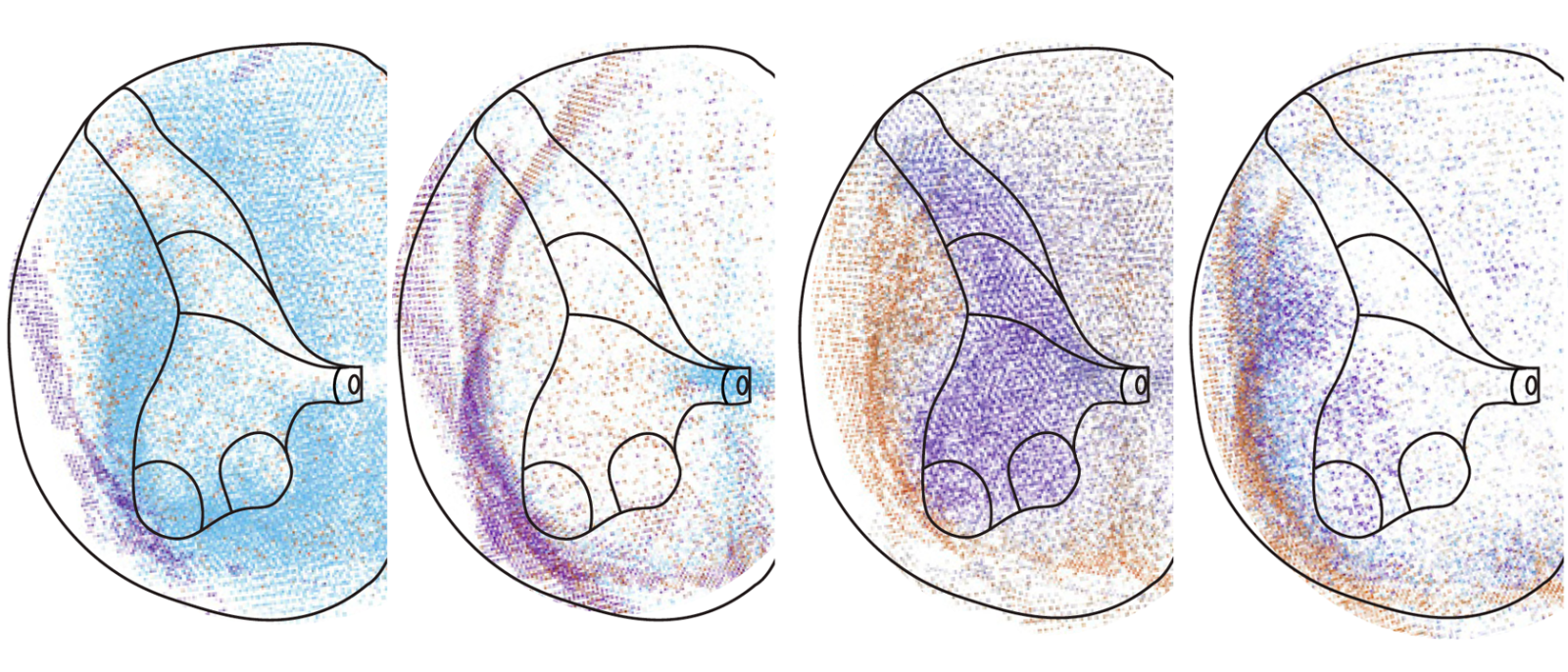
Top image: Different colored dots represent different cells types found in a section of the human spinal cord.