Cells That Expect the Unexpected
Columbia researchers in Professor Rafael Yuste’s lab discover neurons that detect novel stimuli.

We are constantly inundated with sensory stimuli in our environments, amounting to more information than we can efficiently process. But the brain protects us from this onslaught. Decades of research has suggested that our brains automatically suppress responses to predictable stimuli, yet increase responses to novel stimuli. However, this automatic novelty detection is deficient in people with major psychotic disorders such as schizophrenia, apparent even before the first symptomatic episode.
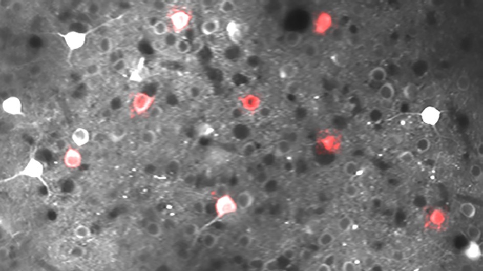
How the brain is able to carry out novelty detection remains a mystery. A group of Columbia neuroscientists in Professor of Biological Sciences Rafael Yuste’s lab recently studied visually evoked brain activity in mice, using similar tests to those used in humans with schizophrenia. When surveying the activity across hundreds of neurons, they found that some neurons on the top layers of the cerebral cortex selectively responded to novel sensory stimuli.
“The notion that these novelty driven brain responses are exhibited in just a subset of the neurons was surprising to us,” said Dr. Jordan Hamm, a former postdoctoral researcher in the Yuste lab and lead author of a study recently published in the Proceedings of the National Academy of Sciences, which documents neurons with these functional properties. “This is a potential game changer for how sensory deficits are studied and understood in psychiatry.”
Selective Groups of Neurons
Earlier research in humans to study how the brain processes novelty has typically involved gross-scale recordings, such as electroencephalography. That work provided a somewhat misleading picture of how the brain actually encodes an unexpected event, suggesting a broad, population-wide response to novel stimuli. The Columbia team now shows that novelty detection is, in fact, carried out by selective groups of neurons.
To make this discovery, the team used two-photon calcium imaging, a type of neuro-imaging that they helped pioneer, which can resolve alive brain activity in an entire region with single-neuron resolution.
“One remarkable thing about these novelty-detecting cells is that they show strong, persistent function connections with each other, forming a specialized novelty-detection ensemble, yet they are mixed in with other cells, doing other things”, said Yuste, senior author of the study.
Cognitive Centers of the Brain Project Predictions
The study also shows that higher cognitive brain regions, such as the prefrontal cortex, send signals backward to dynamically activate or suppress these novelty detecting ensembles. “This suggests a mechanism by which cognitive centers of the brain actively project predictions about the world to lower brain regions, signaling which stimuli to ignore and which stimuli are exciting in the environment,” said Dr. Hamm.
“I also think that, since the ultimate function of the brain could be to predict the future, these novelty detecting neurons could be an essential piece in that puzzle”, said Yuste.
Further study is needed. It will be important to examine cellular, molecular, and genetic properties of these novelty detecting neurons, which could suggest new drugs aimed precisely at ameliorating sensory processing deficits in people with schizophrenia. While the methods do not currently exist to functionally confirm the existence of these cells in humans, pharmacological strategies to selectively augment the function of these neurons could be developed and tested in the future.